Artemis123® Whole Head Neonatal Biomagnetometer
A new, noninvasive investigational tool for pre- and full-term infants
Mapping brain function and detecting neurological abnormalities in infants
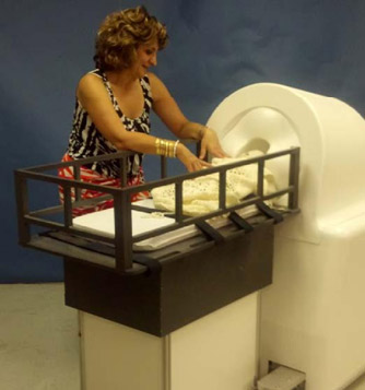
Detection of cortical function in newborns is needed for clinical intervention in the early stages of neurological disorders, before external signs appear and the conditions develop and worsen. Potential uses of Artemis123®,1 for neonatal neurological assessment include:
- Autism
- Epilepsy
- Cerebral palsy
- Perinatal asphyxia
- Hypoxemic-ischemic encephalopathy
- Periventricular white matter injury
- Monitoring recovery from trauma
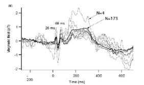
Somatic evoked magnetic field (SEF) obtained from a 7-month old as a function of number of averages from N=4 to 173 epochs. The waveforms are the differences of the SEF at two field extrema. This shows that a small number of averages are needed to acquire SEF data. (data acquired using a Tristan babySQUID® system).
Identifying how infants learn is of interest to many sectors of society, but such studies rely heavily on behavioral analyses. Having a direct measure of cortical activity would provide precise information on the dynamic response in the brain during learning processes. Potential uses of Artemis123® for developmental studies include:
- Mapping of sites and dynamics of sensory functions – auditory, somatosensory, and visual modalities
- Assay stages of nervous system development
1Artemis123® is a registered trademark of Tristan Technologies, Inc. All rights reserved

Unique Features of Artemis123®
- Superior spatial resolution and sensitivity
- Significantly more sensitive to neuronal sources than conventional whole-head MEG systems
- Better spatial resolution compared to existing whole-head MEG sensors
- Better spatial resolution than EEG. EEG signals are distorted by skull discontinuities (fontanels and sutures), making it difficult localize epileptiform tissue.
- Rapid scanning: a typical clinical scan can be completed within thirty minutes
- Anti-vibration construction; infant motion will not cause vibrational artifacts
- Sensor noise level <10 fT/√Hz
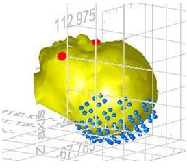
- A dense array of closely-spaced sensors located just below the outer surface of a headrest.
- Allows simultaneous measurement of the occipital area and parietal and temporal areas
- Includes position tracking device and software, permitting measurements during sleep or relatively quiescent wakefulness
- The measurement cradle and companion electronics cart are portable and can be wheeled in and out of elevators, obstetric suites and neonate ICUs
Artemis123® System Description
Principles of Operation
Like adult Magnetoencephalography (MEG) systems, Artemis123® uses superconducting sensors to non-invasively detect and map magnetic fields generated by cortical neural activity. However, Artemis123® takes advantage of the fact that the infant’s scalp and skull are very thin. Tristan’s fabrication methods put the sensing coils very close to the infant brain’s sources of activity, even though SQUIDs must operate in an ultra-cold liquid helium environment. The net result is a significant increase in amplitude of neonate MEG signals. Also, the high density of detectors results in higher spatial resolution compared to adult whole-head MEG.
 System Components
- Bed on mobile cart – easily accessed height
- Power supplies and computer on companion mobile cart to minimize noise
- Subject Tracking – optical tracking system updates movement at 30 Hz with ½ mm accuracy
- Part-wise mapping or optional optical one-click 3D imaging system
SQUID Sensor Array
-
- 606 cm2 sensor coverage area
- 100+ detection coils
- Coil type: 15 mm-diameter first order gradiometers. Adjacent coils can be electronically combined to form planar gradiometers
- Coil gap: ~8 mm from sensor to outer surface
- Gradiometer sensitivity: better than 10 fT/√Hz (referred to proximal windings)
- Reference channels: 12-element reference array for noise reductio
Power and Physical Requirements
• Power: 1.5 kW filtered circuit
• Dewar Cart: 1 m x 2 m x 1.1m (40″ x 79″ x 42″)
• Dewar Cart weight: ~200 kg (~440 lbs)
• Patient Bed weight: <25 kg (<55 lbs)
• Electronics Cart weight: <50 kg (<110 lbs)
Larger coverage areas, higher channel counts, and/or different coil dimensions and configurations are available on request. Contact Tristan for additional information. All Tristan products are covered by a 1-year warranty. Service contracts may be purchased to provide post-warranty coverage.

