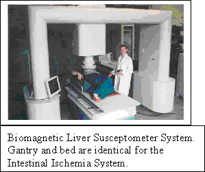
Tristan Technologies fabricates a high sensitivity, multi-channel SQUID magnetometer system for measuring electromagnetic activity in the human intestine. Presently, intestinal ischemia is difficult to diagnose, and is usually fatal. SQUID sensors can detect the magnetic fields produced by the BER (basic electrical rhythm) of the human intestine. The frequency of the BER signals changes under ischemia — the frequency of BER intestinal signals are ~10 cpm (cycles per minute). Magnetic measurements provide improved signal-to-noise

 over the currently more typical cutaneous electrode measurements of electric potential. In contrast to the measurements of voltages on the skin surface, magnetic signals are not attenuated or redirected by the multiple layers of varying electrical resistivity tissues separating the intestine from the skin surface. With multi-channel magnetic measurements, vector projection analysis techniques allow focusing on the signals of interest, distinguishing them from the many other biomagnetic and environmental signals present. Other less serious intestinal disorders, such as Crohn’s disease, ulcerative colitis, and irritable bowel, are also difficult to diagnose; their diagnoses may be improved with this system.
· Non-Invasive — no contact between instrument and abdominal wall.
· Magnetic measurements superior to electric
— signals not attenuated or redirected by the multiple layers of tissue separating skin from intestine
— improved signal-to-noise.
 · Detect signal changes before pathological damage.
· Useful information in short time periods — extensive patient preparation or analysis not required.
Elements in the Tristan Model 637 Intestinal Ischemia System:
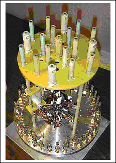
· 29 magnetic field sensing channels, < 20 mm from sensor surface, distributed over
· Large (296 cm2) area of coverage
· Or, intermediate (82 cm2) area of coverage (set at Tristan facility)
· 8 magnetic sensing channels, in a tensor array, monitoring environmental magnetic
noise
· These 37 channels are mounted in the vacuum space of a 27 liter liquid helium dewar
· Helium consumption < 6 liters of liquid per day (volume and consumption allow
system to remain unattended over a long weekend)
· Multi-channel SQUID Control units
· PC computer control for data acquisition and analysis
 The following items are available as part of the system; frequently, users will provide some of these items either to reduce costs or because they have special needs. Tristan is always willing to entertain, and quote on, special requirements.
· Gantry sensor support.
· Patient support, for holding and positioning patient below sensor.
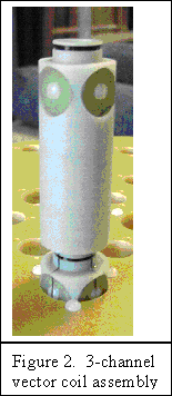
The 29 signal channels are made up of two types of coil assemblies, an axial assembly and a 3-channel vector assembly. The vector assembly is shown in Figure 2. Tthere are 14 axial assemblies and 5 vector assemblies in the system. All the signal channels are first order gradiometers with a 5 cm baseline; the normal (or axial) channel is an axial gradiometer, and, in the vector assemblies, the two transverse channels are planar gradiometers. The gradiometer design assists in reducing environmental magnetic noise, which comes from distant sources and thus couples equal but opposite signals into the two coils making up the gradiometer. Signals from sources of interest are nearby and thus couple much more strongly into the near gradiometer coil; the distant gradiometer coil causing little loss of signal. Thus the gradiometer design only impacts signals when dealing with environmental noise sources; for the ischemia signals of interest, the signal coils can be considered as magnetometers.
 The 14 axial signal channel assemblies measure only the component of the magnetic field normal to the outer surface of the sensor, i.e., e the magnetic field normal to the body of the subject. The 3-channel vector assemblies, on the other hand, will measure all 3 components of the magnetic field, revealing the true vector nature of the magnetic field. The vector information is necessary in revealing signal source locations and true signal magnitudes. It would be optimum to have vector measurements at each of the 19 measurement sites; however, this would increase the number of signal channels and hence the number of SQUIDs and electronic units, and hence the system cost — the supplied configuration provides an acceptable compromise.
The 8-channel tensor array is positioned well above the signal coils. This location for the tensor array is a compromise: the 8-element tensor array should be distant from the signal coils to avoid any detection of signals of interest; and, the array should be close to the signal coils so that the environmental noise detected is representative of the noise picked up by the signal coils.
 Using 3 magnetometers and 5 gradiometers, the 8-element tensor array is capable of measuring the magnetic field and all its first order derivatives at the location of the array. This information is then sufficient to determine the magnetic field value at other locations to the extent that the field is determined by a linear expression. Certainly the environmental magnetic noise signal will have components higher than the first order; but, in the limited volume defined by the 8-element array and the 29 signal coils, this linear approximation to the environmental noise signals should be quite adequate. The signals from the 8 noise measurement channels are used to reduce the noise in the 29 signal channels using post-processing tools. Basically, any signal that is present in both the noise measurement channels and the signal channels is noise, and should be removed. One goal of adding the 8-element tensor array to the sensor is to allow the sensor to operate in an unshielded clinical environment, not necessitating the use of a magnetically shielded enclosure.
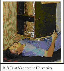
 Analyses of the intestinal ischemia signals are not a part of the system supplied by Tristan. For the details of data analysis, an evolving discipline, we refer you to the group at Vanderbilt University —
www.vanderbilt.edu/lsp/sitemap.htm
References:
Allos, S.H., Staton, D.J., Bradshaw, L.A., Halter, S., Wikswo, Jr., J.P., and Richards, W.O., “Superconducting Quantum Interference Device Magnetometer for Diagnosis of Ischemia Caused by Mesenteric Venous Thrombosis”, World J. Surg. 21, 173–178 (1997)
Bradshaw, L.A., Irinia, A., Sims, J.A., Gallucci, M.R., Palmer, R.L., and Richards, W.O., “Biomagnetic characterization of spatiotemporal parameters of the gastric slow wave”, Neurogastroenterol Motil. 18, 619-631 (2006)
Bradshaw, L.A., “Biomagnetic techniques for assessing gastric and small bowel electrical activity”, in Vargas FM, Franco RH, and Juarez GG., Proceedings of the 8th Mexican Symposium on Medical Physics, 724, 8-13 (2004)
Richards, W.O., Garrard, C.L., Allos, S.H., Bradshaw, L.A., Staton, D.J., and Wikswo, J.P., Jr. Noninvasive diagnosis of mesenteric ischemia using a SQUID magnetometer. Annals of Surgery, 221, 696-705 (1995)


